If the reason lies deeper, then additional research is needed:
- One cat's pupil is big and the other is small. Will it go away on its own? What to do?
- Causes of anisocoria in cats
- Causes of myositis
- Causes of mydriasis
- Diagnosis for anisocoria in cats
- Diagnosis and treatment
- Different-sized pupils: Anisocoria in cats and dogs
- Causes of parasympathetic innervation disorders
- Treatment for impaired parasympathetic innervation
- Pupils of different size: anisocoria in cats and dogs
- Causes of parasympathetic innervation disorders
- Treatment for impaired parasympathetic innervation
- What is anisocoria and what are its causes
- Intoxication
- Nervous system malfunction
- Diagnosis
- One pupil of the cat is larger than the other.What is it? it was fine before!
- What is anisocoria?
- Ophthalmic diseases as a cause of anisocoria
- Different size pupils in a cat – causes
- Anisocoria
- Poisoning, injuries
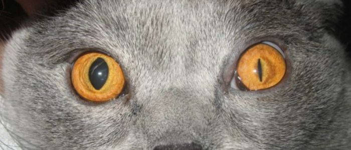
One cat's pupil is big and the other is small. Will it go away on its own? What to do?
Well, first of all, when the eyes are in the dark, they get bigger. If you want to make sure, stand in front of a mirror and close your eye with your hand for a few minutes and see what happens. So if that's the reason, it's not a big deal, the cat just closed one eye or shaded it from the light (by the way they can sleep with their eyes almost open, just a film on top)
This may be the cause; in humans it is called anisocoria or Horner syndrome
Anisocoria means unequal size of the pupils, is not a disease proper, but accompanies another pathology. There are about 30 causes of this phenomenon: corneal trauma, age-related iris changes, adhesions, intraocular pressure changes, nerve impulse transmission disorders, retinal tumors and pathologies. Disorders of the brain and spinal cord can change the size of the pupil. Anisocoria can occur at any age.
The size of the pupil regulates the amount of light that enters the eye. If one pupil is more dilated than the other normal eye, bright light can impair vision to the extent that the light is intense. The animal usually compensates for the excess light intake by partially covering the eye with an eyelid. If the affected eye has a constricted pupil, vision under bright light is normal, but vision is impaired under dim light. Despite the disease, the animals retain good vision, and signs of discomfort are weak or not evident. In some cases, normal pupil function returns with time, even in the absence of treatment. Also in other cases, anisocoria persists for a long time (sometimes for life), which depends on the cause of the condition.
Treatment
Treatment depends on the clinical signs. The reason can be found by examining or other diagnostic methods. In some cases the cause may not be elucidated due to limited or unavailable diagnostic methods, or economic possibilities of the pet owner. Treatment tactics are determined by the medical history, accompanying clinical signs, age and general health of the animal.
Causes of anisocoria in cats
Causes of myositis
- Uveitis is inflammation in the iris and ciliary body (the front part of the vasculature, which works like the membrane in a camera and is responsible for eye color).
- Horner syndrome is the result of interruption of sympathetic innervation of the eye (depending on localization, it can be central, preganglionic and postganglionic).
- Superficial corneal damage – miosis occurs reflexively due to trigeminal nerve irritation, for example, with a foreign object.
Causes of mydriasis
- Medication-induced – caused by medications, such as special drops used at the appointment for diagnosis.
- Iris atrophy – the physiological process of thinning of the iris as the animal ages.
- Glaucoma – a complex of eye diseases that leads to increased intraocular pressure.
- Retinal detachment – a disease in which the retina shifts from its normal position and dies off.
- Optic neuritis – Inflammation of the optic nerve.
- Eye nerve damage – damage to the oculomotor nerve. Cats have two ciliary nerves, the nasal nerve and the malar nerve, so a D-shaped pupil to the right or left side is observed when one of them is affected.
- Central causes (e.g., a neoplasm).
Diagnosis for anisocoria in cats
- collection of anamnesis.
- check the dazzle, pupillary, photochromic reflexes.
- examination with a slit lamp (biomicroscopy).
- Tonometry (measuring intraocular pressure).
- Ophthalmoscopy (examination of the retina with special equipment, an ophthalmoscope).
- If the cornea is damaged, fluorescin staining is performed.
- If necessary, pharmacological tests are performed.
Anisocoria occurs with a large number of diseases, many of which end in blindness and eye removal if not treated in time. When the cat's pupils are of different size the right thing to do is to contact an ophthalmologist at an early date.
Diagnosis and treatment
Sometimes different pupils in a cat appear as a result of a trifling injury or stress. In such situations, the problem disappears within a day. Often the condition of the pet is not a cause for concern. He sleeps peacefully and eats well, there are no problems with toileting. But if the symptoms do not disappear after a while, it is worth suspecting the disease.
Only a doctor can diagnose and determine the root cause of why one pupil is narrow and the other wide, after examination. Veterinarians insist on such procedures:
The disease is quite difficult to treat, so the owner of the pet must be prepared for a long recovery. The rehabilitation of the pet directly depends on the causes of the disease. The doctor may prescribe treatment with antibiotics or anti-inflammatory drugs. The presence of tumors requires surgical intervention. It is necessary to identify and treat the underlying disease and the size of the pupils in both eyes will no longer be a concern.
Different-sized pupils: Anisocoria in cats and dogs
Author: Ekaterina Vasilyeva, veterinary ophthalmologist at the Veterinary Clinic of Neurology, Traumatology and Intensive Care, St. Petersburg, 2017.
Anisocoria – Different pupil size, not uncommon in cats and dogs.
The doctor's first task is to determine which sign is abnormal: constricted or dilated. To do this, observe the pupils when the light is off (a pathologically constricted pupil will not dilate, but will remain narrow). Then the pupil reflex is checked (a pathologically dilated pupil will not respond to light).
Thus, it is determined what we have to deal with: unilateral miosis or mydriasis.
Miosis – A constriction of the pupil, may occur with conditions such as uveitis, superficial corneal defect, and Horner's syndrome.
Uveitis – inflammation of the ocular vasculature, additionally accompanied by symptoms such as turbidity of the intraocular fluid, precipitates on the corneal endothelium, decreased intraocular pressure, and conjunctival hyperemia (Fig. 1). These symptoms help to differentiate uveitis from other diseases. Local and systemic anti-inflammatory therapy is used to treat uveitis.
Miosis in superficial corneal damage occurs reflexively due to irritation of the trigeminal nerve. Superficial damage is also easy to differentiate with a fluorescein test and slit lamp (Fig. 2), and some foreign bodies can be seen even with a flashlight pen. Treatment of these patients will require finding and eliminating the triggering factor (foreign body, lash growth abnormality) and then using a topical antibiotic.
Miosis in Horner syndrome is caused by impaired sympathetic innervation of the iris and is combined with eyelid ptosis, third eyelid protrusion, and enophthalmos (Fig. 3). Visual function is not affected in this syndrome.
Sympathetic innervation of the eye originates in the hypothalamus, where there are neurons of the first order, and their axons go in the spinal cord to the preganglionar neurons located in the first three segments of the thoracic spinal cord. The axons of these preganglionic cells exit the spinal cord and terminate in the cranial cervical ganglion. Here the synapse occurs, the postganglionic fibers leave the ganglion, pass between the tympanic bladder and the stony bone into the middle ear cavity, and go to the eye, where they innervate the pupil dilator muscle (Fig. 4).
Damage to any part of this pathway can cause Horner's syndrome; depending on the location of damage, we distinguish between first-order (hypothalamic – spinal cord), second-order or preganglionic (spinal cord – cervical ganglion) and third-order or postganglionic (cervical ganglion – eye) syndrome.
Causes of parasympathetic innervation disorders
Causes of parasympathetic innervation disorders include inflammation, neoplasm, or trauma in any part of the parasympathetic pathway, such as nerve group damage in cavernous sinus syndrome (mydriasis, ptosis, inability to retract the eye, decreased corneal sensitivity), isolated lesion of the parasympathetic fibers of the glasomotor nerve (internal ophthalmoplegia), complete damage of the glasomotor nerve, including motor parts (external ophthalmoplegia). In cats, a ciliary ganglion lesion in leukemia (FeLV) is considered to cause D-shaped pupil.
Treatment for impaired parasympathetic innervation
Once the lesion is localized, an MRI diagnosis of the selected area can be performed to determine the cause and prognosis, as well as the choice of specific treatment (e.g., removal of a neoplasm).
Anisocoria can occur in many diseases, some of which are ophthalmologic and some neurologic. A thorough examination is required to determine the exact cause of anisocoria and to prescribe treatment.
- Maggs D. J., Miller P. E., Ofri R. Slatter's fundamentals of veterinary ophthalmology 5ed. Elsevier. St. Louis. 2013, 506 p.
- Gelatt K. N. Veterinary Ophthalmology 5ed. Wiley-Blackwell. Ames. 2013, 2170 p.
- Jaggy A., Couteur R. Small animal neurology. Schluetersche. 2010, 528 p.
- Lorenz M. D., Coates J., Kent M. Handbook of veterinary neurology 5 ed. Elsevier. St. Louis. 2010, 560 p.
- Grozdanic S. D., Kecova H., Lazic T. Rapid diagnosis of retina and optic nerve abnormalities in canine patients with and without cataracts using chromatic pupil light reflex testing. Veterinary Ophthalmology. 2012: 1-12.
Pupils of different size: anisocoria in cats and dogs
Author: Ekaterina Vasilyeva, veterinary ophthalmologist at the Veterinary Clinic of Neurology, Traumatology and Intensive Care, St. Petersburg, 2017.
Anisocoria – Different pupil size, not uncommon in cats and dogs.
The doctor's first task is to determine which sign is abnormal: constricted or dilated. To do this, observe the pupils when the light is off (a pathologically constricted pupil will not dilate, but will remain narrow). Then the pupil reflex is checked (a pathologically dilated pupil will not respond to light).
Thus, it is determined what we have to deal with: unilateral miosis or mydriasis.
Myositis – constriction of the pupil, can be seen in conditions such as uveitis, superficial corneal defect, and Horner syndrome.
Uveitis – inflammation of the vasculature of the eye, additionally accompanied by symptoms such as turbidity of the intraocular fluid, precipitates on the corneal endothelium, decreased intraocular pressure, and conjunctival hyperemia (Fig. 1). These symptoms help to differentiate uveitis from other diseases. Local and systemic anti-inflammatory therapy is used to treat uveitis.
Miosis in superficial corneal damage occurs reflexively due to irritation of the trigeminal nerve. Superficial damage is also easy to differentiate with a fluorescein test and slit lamp (Fig. 2), and some foreign bodies can be seen even with a flashlight pen. Treatment of such patients will require finding and eliminating the provoking factor (foreign body, pathology of eyelash growth) and then using a topical antibiotic.
Miosis in Horner syndrome is caused by impaired sympathetic innervation of the iris and is combined with eyelid ptosis, third eyelid protrusion, and enophthalmos (Fig. 3). Visual function is not affected in this syndrome.
Sympathetic innervation of the eye originates in the hypothalamus, where there are neurons of the first order, and their axons go in the spinal cord to the preganglionar neurons located in the first three segments of the thoracic spinal cord. The axons of these preganglionic cells exit the spinal cord and terminate in the cranial cervical ganglion. Here the synapse occurs, the postganglionic fibers leave the ganglion, pass between the tympanic bladder and the stony bone into the middle ear cavity, and go to the eye, where they innervate the pupil dilator muscle (Fig. 4).
Damage to any part of this pathway can cause Horner's syndrome; depending on the location of damage, we distinguish between first-order (hypothalamic – spinal cord), second-order or preganglionic (spinal cord – cervical ganglion) and third-order or postganglionic (cervical ganglion – eye) syndrome.
Causes of parasympathetic innervation disorders
Causes of parasympathetic innervation disorders include inflammation, neoplasm, or trauma in any part of the parasympathetic pathway, such as nerve group damage in cavernous sinus syndrome (mydriasis, ptosis, inability to retract the eye, decreased corneal sensitivity), isolated lesion of the parasympathetic fibers of the glasomotor nerve (internal ophthalmoplegia), complete damage of the glasomotor nerve, including motor parts (external ophthalmoplegia). In cats, a ciliary ganglion lesion in leukemia (FeLV) is considered to cause D-shaped pupil.
Treatment for impaired parasympathetic innervation
Once the lesion is localized, an MRI diagnosis of the selected area can be performed to determine the cause and prognosis, as well as the choice of specific treatment (e.g., removal of a neoplasm).
Anisocoria can occur in many diseases, some of which are ophthalmologic and some neurologic. A thorough examination is required to determine the exact cause of anisocoria and to prescribe treatment.
- Maggs D. J., Miller P. E., Ofri R. Slatter's fundamentals of veterinary ophthalmology 5ed. Elsevier. St. Louis. 2013, 506 p.
- Gelatt K. N. Veterinary Ophthalmology 5ed. Wiley-Blackwell. Ames. 2013, 2170 p.
- Jaggy A., Couteur R. Small animal neurology. Schluetersche. 2010, 528 p.
- Lorenz M. D., Coates J., Kent M. Handbook of veterinary neurology 5 ed. Elsevier. St. Louis. 2010, 560 p.
- Grozdanic S. D., Kecova H., Lazic T. Rapid diagnosis of retina and optic nerve abnormalities in canine patients with and without cataracts using chromatic pupil light reflex testing. Veterinary Ophthalmology. 2012: 1-12.
What is anisocoria and what are its causes
Anisocoria is an alarming symptom that may indicate various diseases in cats. Owners need to consider the high pain threshold of furry pets. They can experience and tolerate pain for long periods of time without showing other signs of serious illness. That's why monitoring cats becomes a very important part of care and concern. If a cat's pupils remain of a different size for a long time, it is necessary to hurry with the diagnosis and prescription
The main difficulty is that anisocoria can be a manifestation of a very wide list of diseases, quite different in the nature of the occurrence and the treatment needed.

Intoxication
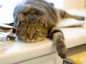
Poisoning can occur when toxic substances are ingested with food. Pets are especially at risk outdoors. Owners know that cats, predators by nature, often taste different kinds of plants. In this way they make up for vitamin and chemical deficiencies. As a rule, their instincts allow them to determine the usefulness of the plant they eat. But sometimes there are exceptions. In this situation, intoxication can occur. Vomiting, lethargy, unsteady gait may be additional symptoms suggesting that everything is not normal with your cat. Similar symptoms can occur as side effects while taking medications.
Nervous system malfunction
Head trauma or concussion received from a fall or a blow can also provoke the manifestation of anisocoria. In this case, the difference in pupil size will be caused by damage to the eye nerve. A more serious cause may be a problem with the cerebellum or cancer. The neoplasms do not allow the eye to fully perform its physiological functions, which leads to these visual manifestations.
Diagnosis
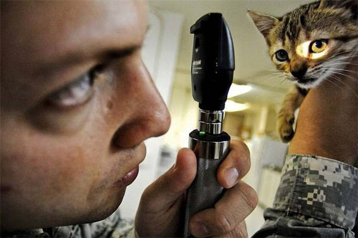
At any veterinary clinic, the doctor will first conduct an interview: the animal's living conditions, chronic diseases, and symptoms that have caused concern. This will be followed by a visual examination. Already with the help of a slit lamp, an experienced specialist will be able to establish the cause of anisocoria, if it is caused by mechanical damage. It is often accompanied by other symptoms: corneal discoloration or clouding, active secretion of lacrimal glands, eyelid disorders.
If the cause is deeper, further investigations are necessary:
In special cases, if the causes cannot be identified by basic diagnostic methods, it is necessary to resort to checking the functioning of the brain with a tomography or magnetic resonance imaging.
One pupil of the cat is larger than the other.What is it? it was fine before!
Anisocoria means unequal size of the pupils, it is not the disease itself, but it is associated with other pathologies. There are about 30 reasons for this phenomenon: traumas of the cornea, age-related changes in the iris, adhesions, changes in intraocular pressure, impaired transmission of the nerve impulse, retinal tumors and pathologies. Disorders of the brain and spinal cord can change the size of the pupil. Anisocoria can occur at any age.
The size of the pupil regulates the amount of light that enters the eye. If one pupil is more dilated than the other normal eye, bright light can impair vision to the extent that the light is intense. The animal usually compensates for the excess light by partially covering the eye with an eyelid. If the affected eye has a constricted pupil, vision in bright light is normal, but vision is impaired under dim light. Despite the disease, animals retain good vision, and signs of discomfort are weak or do not appear. In some cases, normal pupil function returns with time, even in the absence of treatment. Also in other cases, anisocoria persists for a long time (sometimes for life), depending on the cause of the condition.
Treatment depends on the clinical signs. The cause may be found by physical examination or other diagnostic methods. In some cases the cause may not be elucidated due to limited or inaccessible diagnostic methods, or economic possibilities of the pet owner. The treatment tactics are determined by the medical history, accompanying clinical signs, age and general health of the animal.
It's simple. the muscles in the eye have contracted and can't uncompress. go to the vet, they will prescribe you drops. the muscles will relax and everything will be as it was before.
What is anisocoria?
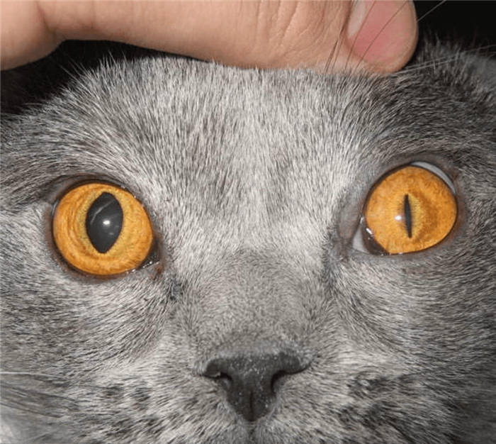
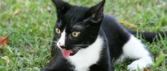
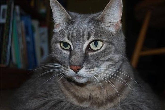
Normally, a cat's pupils are the same. They narrow and dilate absolutely identically. However, sometimes cat owners may notice that one pupil of the cat is larger than the other, or when the light fluctuates, only one eye reacts, while the other remains narrow or wide. Sometimes the changes occur unevenly. What does this sign mean?
This phenomenon is called anisocoria. With anisocoria, the pupils are different in size and respond differently to the amount of light or a stressful situation. Anisocoria itself is not a disease, it only indicates ophthalmologic or other diseases of the animal.
Ophthalmic diseases as a cause of anisocoria
Most often, anisocoria is a consequence of ophthalmologic pathologies. The cause may be:
- Glaucoma. A group of eye diseases characterized by increased intraocular pressure. Visual acuity decreases and the intraocular muscles and optic nerve atrophy.
- Uveitis. Inflammation of the uveal tract, the vasculature of the eye. Typical symptoms are worsening of vision, redness of the sclera, tearing, and constriction of the pupil of the affected eye. Untreated uveitis quickly leads to blindness.
- Scarring between the iris and the lens. Often appear after eye trauma, uveitis, or other ophthalmic diseases.
- Atrophy of the inner eye muscles. In the diseased eye, the pupil is more often dilated. Degenerative changes may be a result of aging or trauma, infections.
- Malignant tumors of the eyeball. Eye cancer is a rare disease, but it can also manifest as anisocoria.
Different size pupils in a cat – causes
Different pupils in a cat – usually a sign of disease, brain vessels or received head trauma. In this case, you need to pay attention to the physical condition of the animal and its behavior.
Heterochromia is a variegated eye. At the same time the iris is not the same. Heterochromia can be complete or partial. In the second case, only part of the eye is colored differently.
If the cat has fallen, the resulting blow to the head may cause a mild cerebral concussion. Even minor vascular damage may provoke dilation of the pupils. Most often, after a minor injury the eyes return to normal within a couple of hours. If this does not happen after a day – you need to take your pet to the vet.
- Unequal pupils in the pet may appear due to a failure of the nervous system. The cat should be given a comfortable quiet corner and, if necessary, medication prescribed by the veterinarian. It can also be caused by old age, inflammation of the eyes, or the appearance of tumors.
- Divergent pupils can appear against the background of untreated cancer. Metastases quickly spread to organs, including the brain. This results in a different pupil constriction effect.
- This effect can be caused by a suffered stroke.
If the cat has one pupil narrow, the other wide – this phenomenon sometimes occurs against a background of stress. Most often, the unusual effect goes away on its own as soon as the animal calms down.
Anisocoria
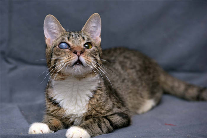
One of the most common feline problems is anisocoria. This is different pupils in size. One of them appears to be underdeveloped. When cats have anisocoria, only one pupil is dilated. It is completely unresponsive to light. Causes of the disease:
Poisoning, injuries
The reason why a cat has different-sized pupils may also become the reason why the animal took some drugs that affect visual functions. The same reaction is sometimes caused by some plants. A healthy eye may not react to mild intoxication, while a sick eye may not, because it is more sensitive.
If the cat staggers in its gait, there is a lack of coordination and vomiting, this is an indication of poisoning. Toxins are the cause of the changes in the eyes. They can enter the animal's body with food or from poisonous plants that the cat has chewed.
When scarring or clouding of varying intensity begins on the eyes after an injury, not enough light enters the animal's eyes. The body's reflex reaction is to dilate the pupil, it becomes larger. So it captures more light. At the same time, the bright reaction is weaker than in the normal state.
Different eyes do not always indicate disease. Cats' pupils change as soon as they smell or see a dog. It is perceived as a clear threat. However, the underlying cause becomes disease.